%20(no%20fill-%20cropped).png)
Using x-ray-vision-like technology to see inside the human brain
- arianacahn
- Dec 1, 2021
- 2 min read

Updated: Dec 21, 2021
Neurons are small. Crazy small. There an estimated 100 billion neurons inside each human brain, so you can probably imagine how tiny these cells are.
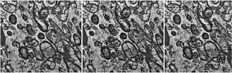
Axon diameter measurements on an image taken using electron microscopy. Taken from Herrera et al. 2022.
As we've learned in a previous blog post, neurons are the primary cells that make up the brain and nervous system. They conduct electrical impulses to get information from one place to another. However, it's not just one long neuron that's running from the top of your head to the tip of your toes. There are many neurons all packed in a nerve together, so that if one or two of the neurons get injured or blocked, there is a redundancy that make sure the information gets where it needs to go!
The same goes for within the brain. Like I said above, there are about 100 billion neurons that make up the human brain, tightly packed in there to make it seem like one big semi-solid structure. Not all neurons within these bundles have the same diameter, though; there is a wide range of microscopic widths possible.
Now here comes the really cool point of this article - University of Winnipeg-affiliated scientists (where the two lead authors were females - so cool!) have used nuclear magnetic resonance (NMR; which is similar to magnetic resonance imaging, or MRI) to measure the tiny widths of these cells. (You can find the original paper here)
Using magnets to peer into live human brains isn't the unusual part of this study - I've done quite a bit of it myself. What the authors did, however, was use a sample of human brain and ran it through a magnet system in order to see and distinguish between (or, 'resolve') some of the smaller neurons in the brain. They were the first to measure these cell widths that were only 2 microns wide. That's 0.0000002 meters (0.002 mm) wide 🤯; which is several times thinner than human hair!


The machine they used looked like the image on the left, whereas an MRI for humans can be seen on the right
The authors of this paper state that these smaller neurons are some of the most important ones, that they are vital to transporting information throughout the brain. They think that it's possible that these neurons are affected or damaged in neurological disorders such as schizophrenia - so using this imaging method, they could potentially diagnose these disorders at an earlier stage, leading to better outcomes for patients in the long run.
This is the first article I've seen in a while that's really got me excited. It's got everything that I need:
Canadian research? check
MRI? check
mental health disorders? check
early diagnostic neural biomarkers? check
female authors? DOUBLE CHECK!
While the work that I've done in the past has looked at collections of neurons in the brain, looking at the individual cells is just as important, if not more, so I'm super excited to see where this research goes in the future!


Comments